ALERT
ALERT ATTENTION ⚠️
In observance of a holiday, Agilent CrossLab/iLab Operations Software Support Help Desk will be closed during U.S. hours on Monday, May 25th, 2026. We will resume regular U.S. support hours on Tuesday, May 26th, 2026. EU and APAC Support will remain open during this time. For urgent matters, please add "Urgent" to the ticket/email subject or press "1" when prompted to escalate a call on the iLab Support phone, and we will prioritize those requests first.
COBRE Integrative Neuroscience
The following cores (BRAIN, CMI, Neuroimaging and VR-AR Core) are supported by the COBRE Integrative Neuroscience (Dr. Mike Webster, PI). Any work done on core instruments and its services must acknowledge the Integrative Neuroscience COBRE P30 GM145646 grant support. A sample statement would be: "Research reported in this [publication, release] was supported by National Institute of General Medical Sciences of the National Institutes of Health under grant number P30 GM145646.”
Cellular and Molecular Imaging (CMI)
The COBRE Cellular and Molecular Imaging (CMI) Core at the University of Nevada, Reno (UNR) housed in the Biology Department provides users with instruments and services for analyzing and quantification of biomolecules, and microscopic investigation of cells, tissues, organs, and small animals with imaging capabilities for brightfield and fluorescent microscopy. The cellular imaging unit of the core maintains several instruments including a Leica SP8 confocal with LIGHTNING capabilities, a Scientifica two-photon microscope, a Leica THUNDER 3D-tissue and Model organism with computational-clearing capabilities. The molecular imaging unit of the core maintains multiple instruments for imaging, analyzing and quantifying DNA, RNA and protein such as a Typhoon Phosphoimager, automated PCR-pipetting robot and Real-Time PCR machines. The tissue culture core unit maintains a state-of the-art space and storage for the culture of insect and mammalian cells with instruments including a WOLF Microfluidic Cell Sorter, Cell Counter and microscope systems. Major equipment (microscopes, cell sorter), training and technical assistance are available on a fee-for-service recharge basis to recover costs for major equipment upkeep and service contracts, technical help, and assisted use for imaging specimens by core staff. Smaller equipment is free of charge. For onboarding, training and assisted use, please contact Core Specialist, Andrew Yanez at ayanez@unr.edu. For general core information and support letters, please contact Core Director, Dr. Alexander van der Linden at avanderlinden@unr.edu.
Equipment and Technology:
Neuroimaging
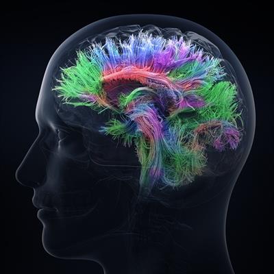
The COBRE Neuroimaging Core at the University of Nevada, Reno (UNR) provides users with resources to measure neural activity in human subjects. Direct neuronal activity measurement is provided by our two high-density, high-frequency electroencephalography (EEG) systems, while measument of higher spatial acuity is provided by our magnetic resonance imaging (MRI) scanners and near-infrared spectroscopy (NIRS) systems. Additional equipment is available for experimental or diagnostic measurement of retinal activity (two ERG systems, including multifocal and visual evoked potentials); behavioral visual function including color blindness, visual acuity, contrast perception; and imaging of retinal structures (AOSLO).
Equipment and Technology:
Virtual and Augmented Reality (VR-AR)
The Virtual and Augmented Reality (VR-AR) Core facility at the University of Nevada, Reno (UNR) is housed in the Department of Computer Science and Engineering in the College of Engineering (COE) and provides state-of-the-art virtual reality (VR) and augmented reality (AR) equipment as well as training and software development services to aid UNR faculty in neuroscience research. Due to its low cost, equipment is available free of charge, but training and software development services will be made available at $25.00 per hour to recover personnel costs. All users are accepted but priority use is given to former and current COBRE investigators. We are mostly familiar with developing in Unity 3D software.
Equipment in the facility includes:
|
Hours Open
|
Hours Staffed
|
Location
|
|
Monday - Friday
|
Monday - Friday
|
Bridge to Research in AI and Neurocomputing (BRAIN) |
| Monday - Friday 9am - 5pm |
Monday - Friday 9am - 5pm |
Bridge to Research in AI and Neurocomputing (BRAIN)
|
|
Monday - Friday
|
Monday - Friday
|
Cellular and Molecular Imaging (CMI) Biology Department, FA 324, 318, 312, 314 1664 North Virginia
|
|
Monday - Friday
|
Monday - Friday
|
Virtual and Augmented Reality (VR-AR) |
|
Monday - Friday Saturday - Sunday |
By appointment |
Siemens Skyra MRI scanner |
|
Monday - Sunday |
Monday - Friday |
Neuroimaging Suite |
| Name | Role | Phone | Location | |
|---|---|---|---|---|
| Alexander van der Linden |
Cellular and Molecular Imaging (CMI) Core Director, COBRE Integrative Neuroscience
|
775-784-6080
|
avanderlinden@unr.edu
|
Fleischmann Life Sciences, room 339
|
| Lars Strother |
Neuroimaging, Director
|
775-784-8678
|
lars@unr.edu
|
Mack Social Science, room 406
|
| Andrew Yanez |
CMI Core Specialist, COBRE Integrative Neuroscience
|
775-444-5333
|
ayanez@unr.edu
|
Fleishmann Life Sciences, room 311H
|
| Alireza Tavakkoli |
Bridge to Research in AI and Neurocomputing (BRAIN)
|
775-682-6842
|
tavakkol@unr.edu
|
WPEB 417
|
| Michael Rudd |
Bridge to Research in AI and Neurocomputing (BRAIN)
|
206-228-5757
|
mrudd@unr.edu
|
EMM 218
|
| Sean O'Neil |
Neuroimaging, Research Manager
|
(775) 784-4711
|
seano@unr.edu
|
EMM 304
|
| Eelke Folmer |
Virtual & Augmented Reality Director
|
7757847592
|
efolmer@unr.edu
|
WPEB 403
|
| Service list |
| ► Cellular and Molecular Imaging (4) | |||
| Name | Description | Price | |
|---|---|---|---|
| CMI - Assisted Use/Training Leica Confocal |
All new users must complete a basic one-on-one training session by our core staff for use of microscopy instruments regardless of background experience. A basic hands-on training involves procedures and basic subjects, but are not limited to, appropriate start-up and shutdown of systems, configurations, generating quality images, z-stacks, and other policies and handling of the instrument.
Training sessions are one-on-one and may depend on the individual needs and the amount of time needed to learn specific techniques. Assisted sessions involve bringing your prepared sample and the core staff will acquire the images for you. Assisted sessions must take place during regular weekday hours.
Please complete the request form for training/assisted use and tell us as much as you can about your experiment. If you are unsure as to which type of microscope is best for your experiment, please complete the consultation request form to schedule a free consultation. Once a request has been approved, a session on the instrument can be scheduled with core staff approval. Time for training is charged at the assisted use rate for that instrument.
|
||
| CMI - Assisted Use/Training Two-Photon |
All new users must complete a basic one-on-one training session by our core staff for use of microscopy instruments regardless of background experience. A basic hands-on training involves procedures and basic subjects, but are not limited to, appropriate start-up and shutdown of systems, configurations, generating quality images, z-stacks, and other policies and handling of the instrument.
Training sessions are one-on-one and may depend on the individual needs and the amount of time needed to learn specific techniques.
Please complete the request form for training/assisted use and tell us as much as you can about your experiment. If you are unsure as to which type of microscope is best for your experiment, please complete the consultation request form to schedule a free consultation. Once a request has been approved, a session on the instrument can be scheduled with core staff approval. Time for training is charged at the assisted use rate for that instrument.
|
||
| CMI - Assisted Use/Training Thunder 3D-Tissue |
All new users must complete a basic one-on-one training session by our core staff for use of microscopy instruments regardless of background experience. A basic hands-on training involves procedures and basic subjects, but are not limited to, appropriate start-up and shutdown of systems, configurations, generating quality images, z-stacks, and other policies and handling of the instrument.
Training sessions are one-on-one and may depend on the individual needs and the amount of time needed to learn specific techniques. Assisted sessions involve bringing your prepared sample and the core staff will acquire the images for you. Assisted sessions must take place during regular weekday hours.
Please complete the request form for training/assisted use, and tell us as much as you can about your experiment. If you are unsure as to which type of microscope is best for your experiment, please complete the consultation request form to schedule a free consultation. Once a request has been approved, a session on the instrument can be scheduled with core staff approval. Time for training is charged at the assisted use rate for that instrument.
|
||
| CMI - Assisted Use/Training Thunder Model Organism |
All new users must complete a basic one-on-one training session by our core staff for use of microscopy instruments regardless of background experience. A basic hands-on training involves procedures and basic subjects, but are not limited to, appropriate start-up and shutdown of systems, configurations, generating quality images, z-stacks, and other policies and handling of the instrument.
Training sessions are one-on-one and may depend on the individual needs and the amount of time needed to learn specific techniques. Assisted sessions involve bringing your prepared sample and the core staff will acquire the images for you. Assisted sessions must take place during regular weekday hours.
Please complete the request form for training/assisted use and tell us as much as you can about your experiment. If you are unsure as to which type of microscope is best for your experiment, please complete the consultation request form to schedule a free consultation. Once a request has been approved, a session on the instrument can be scheduled with core staff approval. Time for training is charged at the assisted use rate for that instrument. |
||
| ► Bridge to Research in AI and Neurocomputing (9) | |||
| Name | Description | Price | |
| BRAIN - AI/ML Deployment - Flat Rate |
Investigators who require novel AI/ML models designed to enhance their research can request a flat rate to gain access to one-on-one dedicated research support. This model is best suited for research that requires the design, implementation, evaluation and deployment of novel AI toolkits for specific target domains. The flat rate provides access to investigators dedicated access to the core staff throughout the lifetime of the projec |
Inquire | |
| BRAIN - AI/ML Design - Flat Rate |
Investigators who require novel AI/ML models designed to enhance their research can request a flat rate to gain access to one-on-one dedicated research support. This model is best suited for research that requires the design, implementation, evaluation and deployment of novel AI toolkits for specific target domains. The flat rate provides access to investigators dedicated access to the core staff throughout the lifetime of the projec |
Inquire | |
| BRAIN - Software Development |
|
Inquire | |
| BRAIN - Statistical Consulting |
Statistical consultation and support to faculty during proposal writing and as key personnel during project execution |
Internal
$50.00
/hr
External $50.00 /hr Commercial/Industry $200.00 /hr |
|
| BRAIN - AI/ML Consultation |
Internal
$2.50
/hr
External $2.50 /hr Commercial/Industry $10.00 /hr |
||
| BRAIN - Statistical Training |
Statistical consultation and support to faculty during proposal writing and as key personnel during project execution |
Internal
$25.00
/hr
External $25.00 /hr Commercial/Industry $100.00 /hr |
|
| BRAIN - HPC Self Use |
Internal
$2.50
/hr
External $2.50 /hr Commercial/Industry $10.00 /hr |
||
| BRAIN - AI/ML Services |
Internal
$50.00
/hr
External $50.00 /hr Commercial/Industry $100.00 /hr |
||
| BRAIN - AI/ML Training |
Internal
$25.00
/hr
External $25.00 /hr Commercial/Industry $100.00 /hr |
||